Main Office: 541-230-1220 | Wildlife Hotline: 541-745-5324
Wildlife Hospital very busy: please call hotline before bringing in an animal
A Most Remarkable Red-tailed Hawk
Stories of wildlife rehabilitation through the lens of veterinary medicine told by Dr. Claire Peterson
In the wildlife rehab world, we often see patients at their very worst. By the time a wild animal is sick or injured enough to be discovered and captured to help it – well, it is often too late. People don’t like to talk about it, but wildlife rehabbers have to deal with a lot of disappointing and heart-breaking cases no matter how talented, skilled, and hardworking they are. I think that’s one reason it’s really important to celebrate our wins – because one of the reasons I love working at Chintimini Wildlife Center is that we give every patient the best medicine and the best chance we can, sometimes even when the odds aren’t in our favor. And it is my pleasure to start sharing these triumphs with you!
Warning: Graphic Content – Please use your discretion before reading the following blog.
Red-tailed Hawk patient #21-500 (our 500th patient of 2021) arrived at Chintimini on May 2nd. I’m going to be completely honest, that when I peeked in the shaded box he was resting in, I was not certain if he was still alive. He was awkwardly on his side with his head down, but once he noticed me he lifted his head and tried to strike out at me with one of his feet. Birds of prey such as hawks rely on their feet as their main weapon and their primary tool to catch prey – being grabbed by one (called “footing”) can cause serious injury if one isn’t wearing the proper protective equipment (mainly, leather gloves). Their taloned feet have a ratchet-like design of tendons, allowing Red-tailed Hawks to squeeze about 250 PSI (pounds per square inch!) and once they’ve grabbed on, it’s hard for them to let go because of these same tendons. This is an advantage for them because if they catch prey like a rabbit that is trying hard to get away, they don’t have to remember to hang on in the ensuing tussle – their feet automatically hang on for them. For those us working directly with wildlife, it does mean we have to take special precautions and training in how we handle our raptor patients.
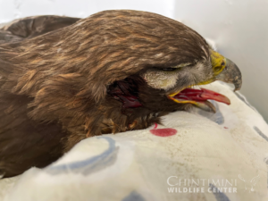
Unfortunately, after being struck by a vehicle and found on the side of I-5 (a major local freeway), #21-500 was in critical condition. He’d been hit hard in the head and was bleeding from his right ear. His right eye was also half-closed and injured, and to top it off he was bleeding from his mouth – a potential sign of internal injuries. We immediately gave him fluid support with vitamins to help his blood pressure from all of the lost blood. We also started him on antibiotics and pain medications for his potential internal injuries. I cleaned up the right side of his head as well as possible but thankfully couldn’t find any obvious skull fractures. The best thing we could do for him now was to give him a warm place and to let him rest – stress in captivity can be a killer, especially to such a debilitated patient.
I was anxious as to how he was doing the next day – those first 24 hours can be critical, especially with internal injuries. He was still alive – a miracle! – but he appeared to be doing worse. He couldn’t stand but instead was laying on his belly in a special “nest” that we make for critical birds so their head is supported up a little and the rest of their body can rest at a comfortable angle. He wasn’t eating on his own but was getting tube-fed some nutrition that he was holding down. I suspected his head trauma had caused swelling around his brain that continued overnight and crossed my fingers the anti-inflammatories we had him on would be enough to help it go down.
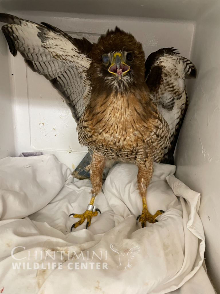
We continued his supportive care and he surprisingly continued to improve. Once he was standing, it was obvious he had opinions about this whole “captivity” thing, and was a true challenge to handle. Due to his aggressive nature, he continued to be a “staff only” patient – to not risk our volunteers’ safety!
As delighted as I was that he was improving, I grew more concerned about his right eye. It often had discharge and seemed dried out – looking closer, it became obvious that although the eye itself was physically fine, the nerves that control the eyelids around it had been injured. He couldn’t blink! Hawks have three eyelids – the upper and lower eyelids like we do, and a third thin eyelid called a “nictitating membrane” that originates from the inner corner of the eye and blinks from inside to outside horizontally. This eyelid acts a little like a goggle – it is mostly clear and allows them to see a bit even if they have to blink while flying, diving, or catching prey. It provides essential protection during these activities while still allowing them to see what they’re doing (see photos below for additional nictitating membrane examples). Unfortunately, since his eye kept drying out, #21-500 kept developing an ulcer on the surface of his eye. After trying several antibiotic drops, ointments, and other things to try to keep it moist, the ulcer was not improving – it only continued to get worse! This can permanently damage the eye, and negatively affect his vision if not corrected quickly.
It was then I decided he needed a drastic measure. Surgery! Not to remove the eye, but to protect it. Under anesthesia, I flushed his right eye with antibiotics, and I sutured his two main eyelids shut over the eye, being very careful to use a technique that didn’t allow the suture material to touch the eye itself or scratch it. Since he couldn’t blink, this would help him keep the eye shut to keep it moist. This was a bit of a scary move for me because if the eyelids were shut with sutures, we couldn’t continue to place antibiotic eye drops in them or monitor how the ulcer was doing. We just had to cross our fingers that the antibiotics, pain medications, and anti-inflammatories he was on would do their work and that the eye would heal in this moist, protected environment.
After a week, it was time to see if this plan had worked. Under anesthesia I removed the sutures holding his eyelids together – I was practically holding my breath – and examined the eye with a special stain to look for ulcers. The ulcer was GONE! It had worked! But now we needed the other piece of the puzzle – would his eyelids regain function to blink as his brain swelling went down? Or were the nerves permanently damaged? Because if he couldn’t regain his ability to blink, that eye would just continually dry out and become injured.
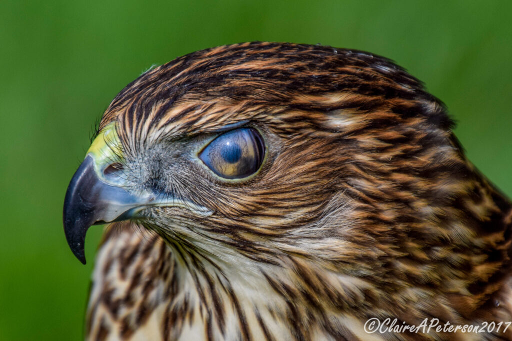
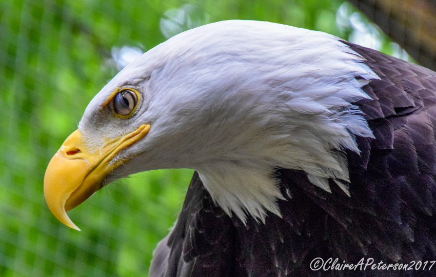
I can’t even tell you the relief I felt when, while he was waking up from the anesthesia, I saw his third eyelid solidly blink across that right eye. Joy! His outer eyelids were still drooping and paralyzed, but if the third eyelid was able to blink, that should be enough to protect his now-healed eye. Now, again, it was a waiting game to see how much he would continue to improve or what would turn out to be permanent damage.
Waiting is so very hard, as #21-500 was in our care for weeks. However, over that time, he slowly gained strength and never seemed to be lacking in coordination despite his head injury. His right eye was still a huge concern to me – but at one of his last rechecks, it appeared he had at least partial control over all three eyelids (If I’m being honest, it’s very difficult to ask a hawk to blink on cue!). #21-500 was flying great, gaining strength, and navigating well in our largest flight cage. It was time for him to go!
Two months later, Red-tailed Hawk patient #21-500 was released back into the wild near where he was found, and I bet you he didn’t turn back once!
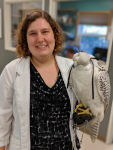
Dr. Claire Peterson
